Biomaterials, Mechanics, & Tissue Engineering
Combining engineering and biology to repair, replace and regenerate organs and tissues
Our students learn to
Conduct interdisciplinary research employing technologies that combine the principles of life science, biomaterial engineering, and clinical applications
Restore, maintain, and improve tissue functions following damage due to disease or trauma
Develop new techniques and biomaterials to guide cell behavior and reconstruct damaged tissues and organs

Utilizing genetically modified mice with temporally activated fluorescent proteins within specific cell types, our laboratory can uncover cell populations (red) that contribute to the formation of diseases such as heterotopic ossification

A 3-D model of ocular wound healing reveals how cells (green) compact and align extracellular collagen (red) through the generation of mechanical forces
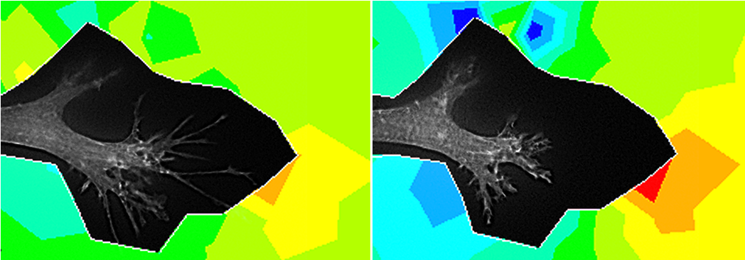
Combining live cell imaging of fluorescently labeled cells with finite element modeling leads to novel insights into sub-cellular patterns of force generation, which can improve our understanding of fundamental processes such as wound healing and embryonic development

Combinations of advanced techniques such as multiphoton fluorescence and second harmonic generation imaging are used to study tissue remodeling and regeneration. Understanding these processes is critical to developing new therapies for treating blindness due to corneal scarring
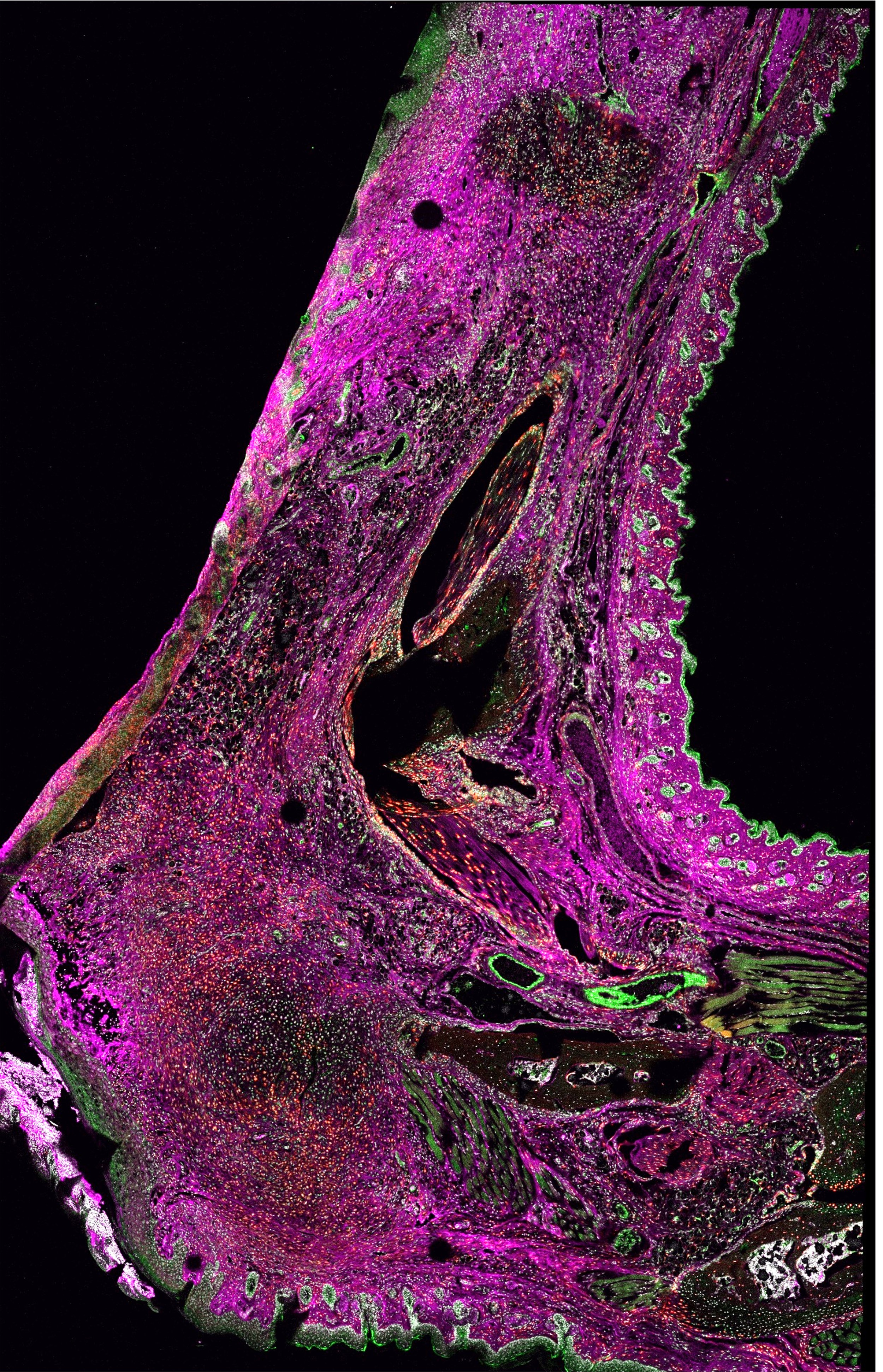
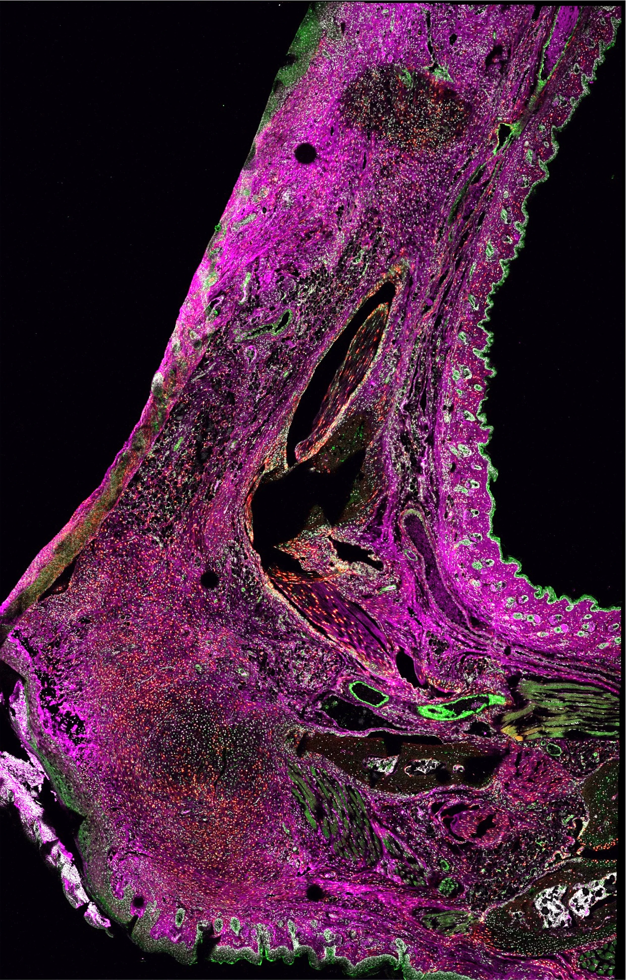
Scanning confocal microscopy imaging allows for whole-section, high-resolution imaging to elucidate the spatial expression patterns of proteins of interest (purple).
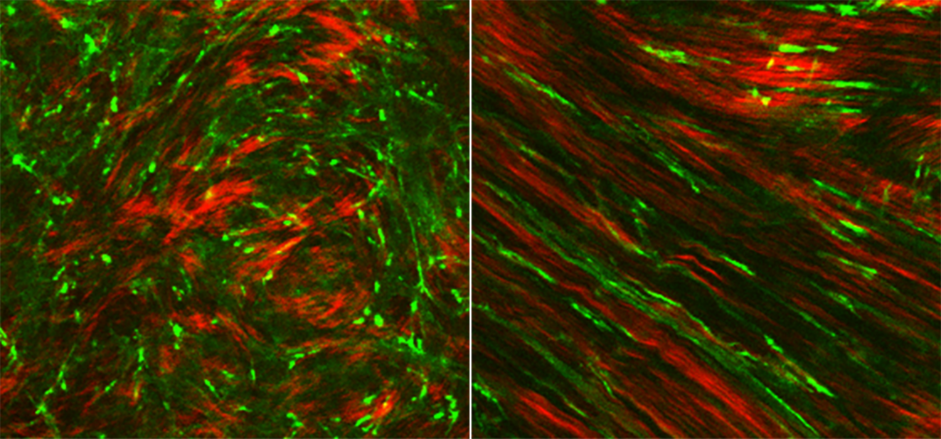
Interactions between cells (green) and the extracellular matrix (red) following laser refractive surgery in the cornea provides insights into how healing and remodeling are regulated
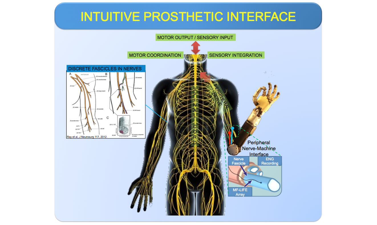
Building intuitive prosthetic interface for seamless motor coordination in amputee patients
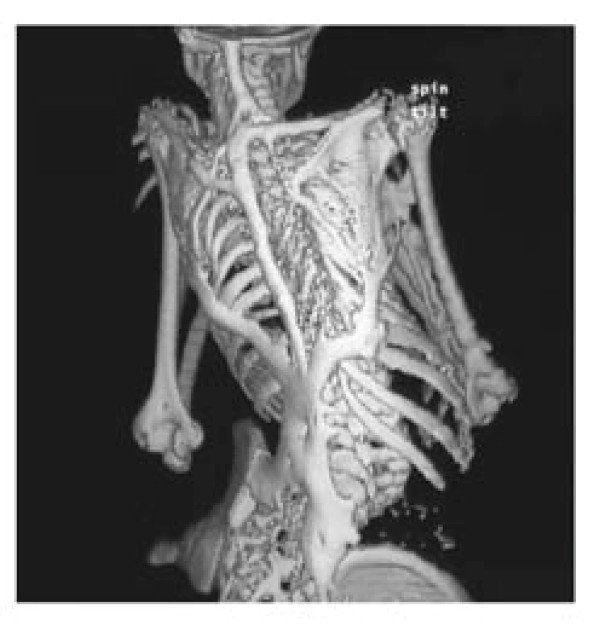
CT scan of human Heterotopic ossification. This is a disorder where the formation of ectopic bone happens in soft tissues of the musculoskeletal system due to an activating point mutation in ACVR1 gene, high energy traumatic injuries such as burn or blast wounds, invasive orthopedic cases, or severe brain and spinal cord injuries
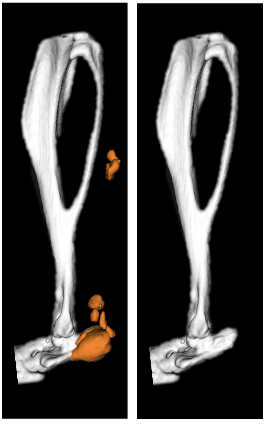
Mouse hindlimb uCT: Levi Lab has generated a mouse model that reliably and reproducibly replicates the formation of human traumatic heterotopic ossification allowing for in vivo studies of the debilitating disease

The Mahendroo Lab uses the mouse as a model system to study term and preterm birth. The focus is to define the molecular steps that transition the cervix from a closed rigid structure to one that opens for a term birth.